
Cone Beam CT (3D) Imaging
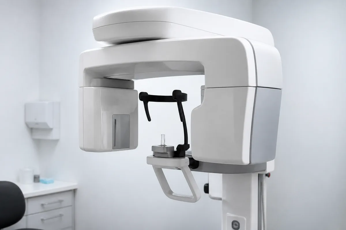
Cone Beam CT (3D) Imaging
Cone beam CT, often called 3D imaging, captures a detailed three-dimensional view of your teeth, jaw, and surrounding bone. Unlike a standard two-dimensional X-ray, it allows a clearer look at structures from multiple angles, which is especially useful when treatment planning calls for extra precision.
When 3D Imaging Is Typically Used
- Dental Implants — Evaluating bone density and position before placement.
- Root Canal Treatment — Identifying complex canal anatomy that isn't always visible on flat X-rays.
- Oral Surgery — Mapping nerves, sinuses, and surrounding structures ahead of extractions or other procedures.
Working Alongside Other Digital Tools
Cone beam CT often pairs well with other imaging already used in general dentistry. Together with digital X-rays for routine diagnosis and a digital impression scanner for restorations, this technology helps build a fuller picture of your oral health so treatment can be planned thoughtfully from the start.
What to Expect
The scan itself is quick and non-invasive, similar in feel to a standard dental X-ray but capturing far more detail in a single pass.
This content is for general information only and isn't dental or medical advice. Talk with a qualified provider about your own situation. If you have a medical emergency, call 911.